Why Therapies for Traumatic Brain Injury Should and Could, Start in the first 5 minutes of Injury
PREAMBLE: Academia has struggled to move the needle against Traumatic Brain Injury (TBI). Daniel Spaite MD (2020 lead author of the largest study of TBI to date – The EPIC Project) opines that
“due to the younger age and ability to survive the insult, TBI causes more suffering and cost to mankind than all Cancers, Strokes, and Heart Attacks… combined.”
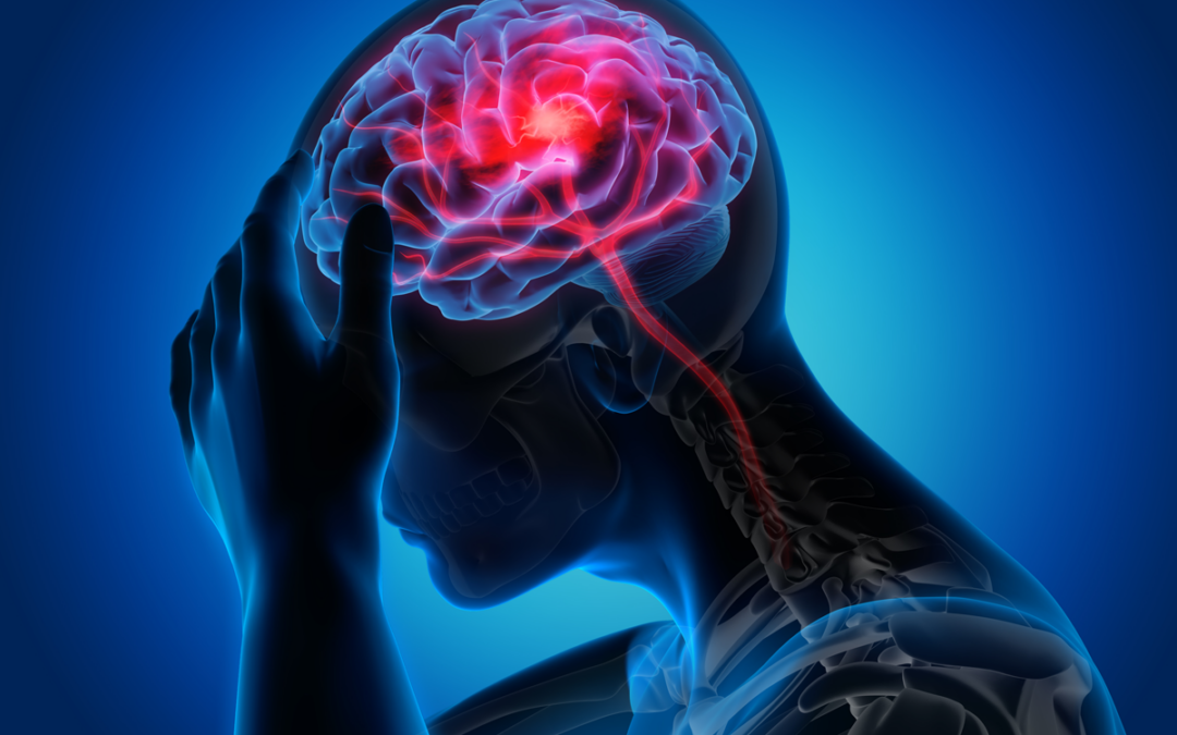
Other than the recent FDA authorization of the Q-Collar™, treatments to “prevent” TBI simply don’t seem to exist. Academic and military research protocols impart a 30-minute post-TBI time point for intervention, as it may take this long for the first responder to arrive to the scene ready to act. Naturally, the treatment challenge against the known tsunami of inflammatory and oxidative chemical cascades already lags because the damage already started within the first five minutes of injury. See Figure 1. Historically, The GOLDEN HOUR (described in military major trauma and hemostasis care) describes the typical lead time for battlefield therapeutic intervention to begin since rapid transport to primary and secondary care centers allows for more directed care, where the “luxuries” of having diagnostic and other therapeutic modalities exist. Given that much of the inflammatory and oxidative cascades have come and gone in the first five minutes post-injury, there seems to be merit in seeking to accelerate the intervention time to quell the early events leading to such known devastation.

Figure 1 Time course of TBI
In STROKE management, “Time is Brain” was coined by Camilo Gomez in 1993, and the idea of “Brain Attacks” also took hold to help society understand the seriousness of delaying directed therapy in strokes. Conceptually, it is felt that “1.9 million brain cells are lost for each minute of delay of definitive care during the brain hypoxia of stroke.” We contend a similar societal and TBI Community establish a new mindset to adequately accept the challenge of truly altering the scourge of this disease. Operationally, a full 30 minutes after a TBI insult represents the norm for studying therapies. Given the above 5-minute time course, we require radical thinking to provide interventions to the soldiers (or in society) to ensure that we are maximizing brain cell protection immediately after it occurs. Our DeltaChase company continues developing a patented technology called The SAGE Rebreather™, a specialty CO2 Rebreathing device that is incredibly lightweight, collapsible, and essentially failsafe in its ability to deliver designed levels of therapeutic CO2 to the battle or sporting field arena.
DeltaChase discovered how rebreathing “columns of air” gives many animals in Nature control over their body’s CO2 levels. Due to its portability and ease of use, we contend this could become the standard of care to administer life and brain-saving interventions immediately before and after TBI. See FIG 2. These clever little devices could become standard Emergency Medical Technician (EMT) equipment or be carried into conflicts or onto the field athletic endeavors. Full Tech Brief available upon request.

FIG 2. The SAGE COMBAT RECYCLER
SETTING THE STAGE FOR DISCUSSION: David Smith, MD, evolved three theories pertaining to the mechanisms and potential to mitigate TBI based on energy absorption into human tissues and liquids in moving containers. Hydrodynamics (the study of fluids in moving containers, coined by NASA as “SLOSH”) and Cavitation are at the core of these theories, and Smith labeled them as Macro-, Micro-, and Molecular-SLOSH.
Macro-SLOSH
Hypercarbia overwhelmingly represents the strongest determinant of Cerebral Blood Flow, which we anticipate to fully “fill the compensatory reserve volume (CRV)” from an arterial standpoint in a manner similar to the way the Q-Collar’s jugular compression fills CRV from a venous standpoint. We contend, therefore, that all TBI mitigating references for the Q-Collar (roughly 25) should be fully correlative to CO2 Rebreathing modalities.
In the regulatory pathway at the FDA, concerns arose about the potential of increased bleeding when cranial veins were more completely filled up. In response, we performed a study of controlled cortical impact (CCI) on 12 large swine when jugular compression had been imparted. Counter to the FDA’s concerns, we predicted the Q-Collar would substantially REDUCE total hemorrhaging if worn before a penetrating or severe TBI (with resultant torn vessels etc.) We theorized that with all the venous vessels maximally “filled” and with little or no CRV volume left, where would the bleeding go? Our studies showed our jugular compression device (the Q-Collar™) increases the volume within the cranial sinuses between 5-25%. Figure 3 nearby (Published in electronic abstract form)

(Figure 3)
“Filling of the Compensatory Reserve Volume” (roughly 4ml intracranial and 25ml in spinal vertebral space)
More importantly, our published CCI study did show a ~50% improvement in bleeding (by subarachnoid and Intraparenchymal hemorrhage scores).1 The mechanism of therapeutic benefit was thought to be similar to tamponade, defined “as the closure or blockage (of a wound or body cavity) by, or as if by, a tampon, especially to stop bleeding.” Applying compression to bleeding external wounds would accomplish the same result. We anticipate CO2, in the setting of TBI mitigation, should carry all the benefits of previously studied jugular compression and Macro-SLOSH in energy absorption AND the tamponade of potentially bleeding vessels (to reduced risk of intracranial bleeding).
Micro-SLOSH
Smith then began bringing his second theory (Micro-SLOSH) to light, which isn’t actually a new theory, but rather, a “new look” at resurrecting Cavitation Theory (circa the 1950s). In “non-blast induced TBI,” cavitation theory has lost favor (in the minds of academics) in deference to Shear-Strain Theory, which represents the widely-held mechanistic theory of mild TBI to date. Smith contended that conventional wisdom (and literally, the entire TBI scientific community) had been led astray due to overlooking the effects of the Non-Newtonian (shear thinning) aspects of macro- and micro-blood, and thus, the oversimplification of creating spurious cavitation models in the studying of TBI. Only PARTIALLY-FILLED containers can allow fluids to separate and thus form the vapor cavities needed to cavitate.
Sadly, researchers chose to model the cavitation chambers by FULLY-FILLING them to the top, thereby making cavitation more difficult, if not impossible. Further, to improve visibility of bubble formation and collapse, no model was done with actual blood, which is Non-Newtonian (shear-thinning)— thusly being more likely to cavitate. Smith presented his paper on “Cranial Venous Blood Cavitation: A Possible mechanism of Traumatic Brain Injury associated with Sport-Related Head Impacts” at the 2016 San Diego Congress of Neurosurgeon’s annual meeting with Julian Bailes MD (of the movie CONCUSSION fame). The talk was entitled “The Physics of Traumatic Brain Injury-Revisited,” by Smith/Bailes, and it was well received.
It was Geoffrey Ling, MD, PhD, (the US Army’s premier subject matter expert on TBI) that opined, “how does the physics of an impact lead to the chemistry of a TBI?” We believe that cavitation is the only naturally occurring phenomenon that could possibly explain such a dramatic “chemical reactivity.” Cavitation involves fluid mechanics leading to vapor cavities that expand into bubbles and then, upon collapse, release enormous energies, including heat levels approaching those of the sun (9,900°F), 1000 atmospheres of pressure, and together, resulting in a One-MILLION-fold increase in chemical reactivity.
We have demonstrated and published that if you fully fill a container with blood and impact it at nearly any force (IED levels studied), it will greatly resist cavitating and lysing. Both jugular compression and CO2 Rebreathing fill the cranial vault more completely, thus resisting cavitation.
Molecular-SLOSH
So, for a mechanistic explanation of Molecular-SLOSH, Smith says, we “follow the physical energies” and where they can be absorbed into “sloshable” liquid or floppy objects (see the physics concept of elastic and inelastic collisions). Smith contends, “it is all about blood; the brain just may be an innocent bystander.” Poised to absorb energy by hydrodynamic SLOSH mechanisms, we have 80 trillion floppy, overly-compliant, partially-filled (~60%) Red Blood Cells. Inside each RBC is 250 million molecules of floppy-globular hemoglobin (in fact, the RBC’s dry weight is 97% hemoglobin). This all equates to 1.2 x 1024 molecules of floppy, energy-absorbent hemoglobin molecules, all of which are circulating through the brain at a disproportionately high rate compared to the rest of the body (the brain is ~3% of body weight but receives 20% of cardiac output). Within each hemoglobin macromolecule are FOUR HEME POCKETS and (4) IRON (Fe++) molecules, which perhaps represent the body’s most curious and physiologically complex component.
To life, iron is nearly ubiquitous (a few rare forms of life can use copper instead of iron), and in its elemental form, IRON is extremely volatile. So, Nature had to delicately encapsulate iron into large floppy-globular proteins called “The Heme Pockets.” physics tells us these “pockets” represent innumerable pockets for cavitation to occur. Notably, due to the complexity and critical allosteric nature of this heme protein, there is nearly 97% homology of Hgb’s structure within the animal kingdom (i.e., our Hgb is 97% identical to virtually all other forms). Just as amazingly, RBCs have just a 120-day life cycle and thus must be recycled through the highly complex labyrinth of the Porphobilinogen Pathway such that “free Iron” can NEVER be in the direct presence of water and salts. This combination is literally explosive. See Figure 4 nearby. A cavitation blast wave should propagate through blood more readily because the blood not only has large levels of dissolved gases but 4.8 x 1024 heme pockets to initiate further cavitation (four heme pockets per Hgb molecule). We contend that THIS cavitation wave literally opens the heme pockets and allows the iron to react explosively with free water and salts. As a result of this energy release, just ONE electron transfers from oxyhemoglobin to O2, resulting in methemoglobin and superoxide O2 (interestingly, two KNOWN byproducts of TBI). Since methemoglobin cannot carry/deliver any oxygen, the oxidative and inflammatory cascade of events begins to devastate the neuron’s polarity and ability to create energy (ATP). As methemoglobin increases and RBC lysis begins, methemoglobin reductase (the body’s antidote to methemoglobin) can no longer function as this enzymatic reaction can ONLY take place on the inside membrane of INTACT RBCs.

Figure 4. The Heme Pocket
Abating the inflammatory/oxidative/Spreading depolarization cascade “immediately” post-TBI should be dramatic with CO2 ALONE. CO2 could quite honestly be the antidote for all forms of SLOSH (macro, micro and molecular)
-
-
- Assuming the CO2 of the subject is not already, an added 4-6 mmHg ETCO2 should:
-
-
-
-
-
- increase CBF by ~50-75%
- increase intracranial blood volume by ~4ml
- take up the Intracranial Reserve Volume (reducing brain slosh)-and invoke a kind of “tamponade” (note ~25ml increase in spinal vertebral blood volume too)
- shift CSF out of the intracranial space (into peri-spinal space)
- increase dissolved CO2 and HCO3– at the peri-neural space
- increase delivery of oxygen to cerebral tissues
- reduce oxidative damage at the cellular and mitochondrial level
- reduce free radicals (and active oxygen forms), and protect superoxide dismutase against oxidative damage
- reduce blood and CSF pH (HCO3 represents a weak acid)
- reduce neuron excitability by delivering more HCO3 to the neurons
- modulate Ca++ channels and resultant inhibition of Kv7/KCNQ channel activity
- potentially reverse Spreading Depolarizations (a hallmark of neural cell death)
-
-
-
- The topic of Brain stem involvement in TBI causing an alteration in respirations:
- The medulla detects CO2 in the blood and adjusts the frequency of breathing, while the pons controls how long one’s inhale lasts (i.e., tidal volume) by sensing how widely the lungs stretch.
- If the brainstem becomes injured, one might be expected to develop hypercarbia.
- If no brainstem involvement, one might more likely to develop hypocarbia (see below)
- Although difficult to prove a negative, we are struggling to find data showing that pCO2 ELEVATES upon presentation of a TBI victim. In animal studies, hypercapnia is certainly the norm post-TBI. We contend this deserves study.
- The medulla detects CO2 in the blood and adjusts the frequency of breathing, while the pons controls how long one’s inhale lasts (i.e., tidal volume) by sensing how widely the lungs stretch.
- Sympathetic Storm (an argument for the pCO2 to be low, not high, post-TBI)
-
- There is an immediate [within seconds] sympathetic discharge when a TBI injury occurs, raising plasma adrenaline levels to approximately 1,200 times the normal value. The adrenaline levels then fall, but they remain 3 times higher than normal for approximately ten days.[6][7]
- Therefore, one would “expect” a sympathetic outpouring immediately post-TBI to result in tachypnea (shallow breathing RR>40) and, thus, hypocarbia. Again, we emphasize as THEORY, not fact.
- About 25 percent of patients with brain contusions or hematomas, and about 50 percent of patients with penetrating head injuries, will develop immediate seizures that occur within the first 24 hours of the injury.
- We KNOW Hypercarbia treatment breaks seizures.,,
- In TBI, common clinical experience reflects that lesions, i.e., contusions, evolve over the first 3–5 days when edema accumulates and ICP rises.
- “3-5 days”? One would NOT expect a significant rise in ICP in the FIRST 5-10 minutes post-TBI and not even likely within the first 60 minutes of mild hypercarbia (within the Golden Hour). Even extreme hypercapnia should not be detrimental, and even small amounts of hypercapnia SHOULD result in considerable positives.
In Conclusion
The medical community has considered stroke management timing of such paramount importance that it has resolved to call strokes “Brain Attacks” in order to impart a sense of urgency in getting patients to care and starting that care. But, there has NOT been the same urgency to get TBI patients to care as there just wasn’t anything to offer them even IF they arrived within the “Critical Time Period” of the first ten minutes. Now there is a management opportunity that is deployable in the first 10-30 minutes, and it needs to play out in the scientific circles to gather data for or against its use.